tree in bud opacities seen in
However to our knowledge the relative frequencies of. 1 day agoThe centrilobular nodules and tree-in-bud pattern of opacities seen on this patients imaging studies are consistent with aspiration.
2
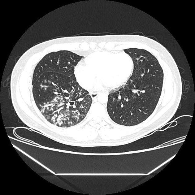
Bronchiolitis is characterized at thin-section CT by the presence of centrilobular nodules and linear branching opacities producing a tree-in-bud appearance Fig 7 4.

. High-resolution CT scan. The differential for this finding includes malignant and inflammatory etiologies either infectious or sterile. The tree-in-bud sign indicates bronchiolar luminal impaction with mucus pus or fluid causing normally invisible peripheral airways to become visible 80.
Tree in bud opacification refers to a sign on chest CT where small centrilobular nodules and corresponding small branches simulate the appearance of the end of a branch belonging to a tree that is in bud. These nodules are centered within the secondary pulmonary lobule without involvement of the subpleural lung compatible with a centrilobular distribution. 3 2005 Tumor emboli from Ewing sarcoma in a 16-year-old boy.
The localized tree-in-bud sign is typically seen in infectious bronchiolitis aspiration bronchiolitis and bronchopneumonia including tuberculosis and nontuberculous mycobacterial pulmonary disease. In addition the centrilobular nodules have a branching configuration and appear to arise from a stalk otherwise known as a tree-in-bud pattern. We aimed to establish the incidence of the TIB pattern as a proportion of all patients undergoing chest CT.
Rossi SE et al Tree-in-Bud Pattern at Thin-Section CT of the Lungs. Bronchiectasis which may be of any cause can produce the tree-in-bud pattern. Its microbiologic significance has not been systematically evaluated.
Tree-in-bud TIB opacities are a common imaging finding on thoracic CT scan. In radiology the tree-in-bud sign is a finding on a CT scan that indicates some degree of airway obstruction. Vascular tree-in-bud sign can be observed in foreign-body-induced necrotizing pulmonary vasculitis cellulose and talc granulomatosis 2 4.
Found that the tree-in-bud pattern was seen in 256 of the CT scans in patients with bronchiectasis. Respiratory infections 119 of 166 72 with mycobacteria 65 of 166 39 bacteria 44 of 166 27 viruses four of 166 3 or multiple organisms six of 166 4 were most common. However to our knowledge the relative frequencies of the causes have not been evaluated.
What causes tree in bud opacities. The tree-in-bud sign is a nonspecific imaging finding that implies impaction within bronchioles the smallest airway passages in the lung. Chest x-ray in a 60 year old patient of Asian extraction demonstrates faint reticulonodular opacities.
The tree-in-bud pattern is commonly seen at thin. The purpose of this study was to determine the relative frequency of causes of TIB opacities and identify patterns of disease associated with TIB opacities. Originally reported in cases of endobronchial spread of Mycobacterium tuberculosis this.
TIB opacities were seen in association with bronchiectasis in 30 123 of 406 of cases and with an apical-predominant disease in 25 10 of 406 cases. TIB opacities can be seen in isolation bronchiolitis 1314 or with other infl ammatory imaging fi ndings includ-. Tree-in-bud opacities appear as tiny centrilobular branching structures on CT most often in.
Tree-in-bud pattern seen on high-resolution CT HRCT indicates dilatation of bronchioles and their filling by mucus pus or fluid. Multiple causes for tree-in-bud TIB opacities have been reported. The tree-in-bud pattern seen on CT represents radiologic sequelae of an infectious or inflammatory process.
Radiologic-Pathologic Overview RadioGraphics Vol. Miller Jr W T Panosian J S. What does tree-in-bud opacities mean.
This is consistent with intrabronchial tumor. Usually somewhat nodular in appearance the tree-in-bud pattern is generally most pronounced in the lung periphery and associated with abnormalities of the. High-resolution CT scans show enlarged and beaded subsegmental arteries in the lower lobes.
Originally and still often thought to be specific to endobronchial Tb the sign is actually non-specific and is the manifestation of. Causes and Imaging Patterns of Tree-in-Bud Opacities. In the remaining 67 273 of.
Causes for TIB opacities were established in 166 of 406 409 cases. In the December 2009 issue of the AJR. Note the peripheral tree-in-bud opacities.
Generally these often result in bronchial wall thickening with replacement of the normally air-filled lumen with mucous or pus. However to our knowledge the relative frequencies of the causes have not been evaluated. 1 refers to a pattern seen on thin-section chest CT in which centrilobular bronchial dilatation and filling by mucus pus or fluid resembles a budding tree Fig.
TIB opacities represent a normally invisible branches of the bronchiole tree 1 mm in diameter that are severely impacted with mucous pus or fluid with resultant dilatation and budding of the terminal bronchioles 2 mm in diameter 1 photo. Multiple causes for tree-in-bud TIB opacities have been reported. TIB opacities were seen in association with bronchiectasis in 30 123 of 406 of cases and with an apical-predominant disease in 25 10 of 406 cases.
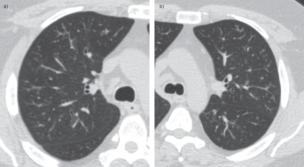
Although seen in various disease conditions their presence usually indicates infectious or inflammatory small airways disease such as by hypersensitivity pneumonitis respiratory smoking-related bronchiolitis and endobronchial spread of infection. CT confims numerous centrilobular nodules with opacified distal bronchioles tree-in-bud sign and bronchiectasis. Patient does have a large para-esophageal lymph node as.
Tree-in-bud TIB is a radiologic pattern seen on high-resolution chest CT reflecting bronchiolar mucoid impaction occasionally with additional involvement of adjacent alveoli. This is very hard to pick up on imaging or prove histologically as this is well beyond the range of bronchoscopy. It is not specific for a single disease entity but is a direct sign of various diseases of the peripheral airways and an indirect sign of bronchiolar diseases such as air trapping or sub.
In radiology the tree-in-bud sign is a finding on a CT scan that indicates some degree of airway obstruction. It consists of small centrilobular nodules of soft-tissue attenuation connected to multiple branching linear structures of similar caliber that originate from a single stalk. The tree-in-bud sign is a nonspecific imaging finding that implies impaction within bronchioles the smallest airway passages in the lung.
Turn on spectral and notice that the tree-in-bud opacities have clear iodine uptake. These findings most likely represents pulmonary TB or MAC despite negative induced sputum specimens. The tree-in-bud pattern is commonly seen at thin-section computed tomography CT of the lungs.
Although initially described in 1993 as a thin-section chest CT finding in active tuberculosis TIB opacities are by. Multiple causes for tree-in-bud TIB opacities have been reported. Patient does not complain of cough.
In the remaining 67 273 of 406 of cases they were seen without permanent structural changes. Thus the bronchioles resemble a branching or budding tree and are usually somewhat nodular in appearance This morphologic pattern can be seen in a wide variety of diseases as illustrated by Gosset et al.
2

Computed Tomography Of The Chest Showed Nodular Opacities With Tree In Download Scientific Diagram

Hrct Scan Of The Chest Showing Diffuse Micronodules And Tree In Bud Download Scientific Diagram

Tree In Bud Pattern Radiology Case Radiopaedia Org
2

Scielo Brasil Tree In Bud Pattern Tree In Bud Pattern

Tree In Bud Sign Lung Radiology Reference Article Radiopaedia Org
2
Tree In Bud Appearance Radiology Case Radiopaedia Org
Hypereosinophilic Obliterative Bronchiolitis A Distinct Unrecognised Syndrome European Respiratory Society

Tree In Bud Pattern Pulmonary Tb Eurorad
Pdf Tree In Bud
View Of Tree In Bud The Southwest Respiratory And Critical Care Chronicles

Tree In Bud Sign Lung Radiology Reference Article Radiopaedia Org
2

Chest Ct With Multifocal Tree In Bud Opacities Diffuse Bronchiectasis Download Scientific Diagram

Tree In Bud Pattern Pulmonary Tb Eurorad

Tree In Bud Pattern Radiology Case Radiopaedia Org

Pdf Tree In Bud Semantic Scholar